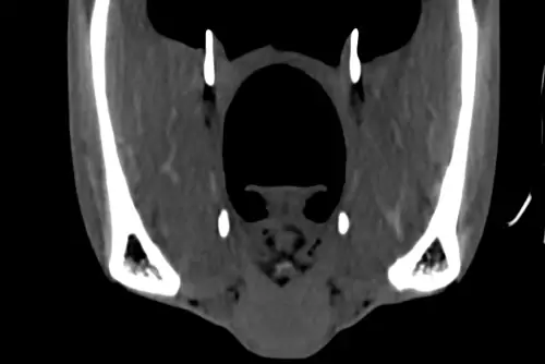
Same horse, same day ... so what have we done to make the CT scan on the left (image 1) look different from the middle one (image 2)?
We have injected contrast into the veins which we can see highlighting the blood vessels (white arrow heads) and the pituitary gland (black arrow) at the base of the brain (image 3).
Contrast enhanced CT is useful for assessing regions of increased or decreased blood flow or leaking blood vessels. This helps us identify soft tissue lesions and infectious or inflammatory processes. The use of contrast enhanced CT imaging is a huge asset to us at BELL EQUINE to improve diagnosis using our state-of-the-art CT scanner and can be performed in standing horses.
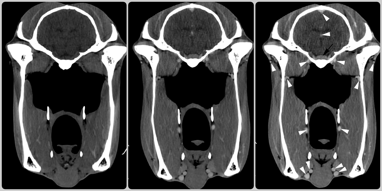
(Image 1: pre-contrast; image 2: post contrast; image 3: marked up image showing contrast uptake)