
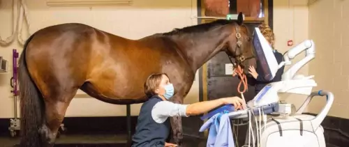
Diagnostic Imaging
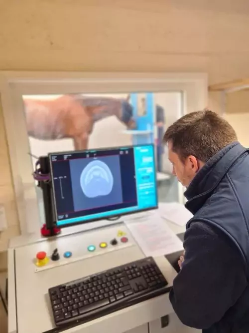
Diagnostic Imaging
Diagnostic imaging is an essential component of the diagnostic work-up of many cases. In many instances, a combination of imaging techniques is required. We have available a range of up-to-date imaging equipment including x-ray generators and digital radiograpgy equipment, a number of different ultrasound machines, a variety of video-endoscopes, a gamma camera (for nuclear scintigraphy), CT (Computed Tomography) and MRI systems. In addition to our first class equipment, we have a number of experienced specialists in imaging that help with the interpretation of the images we obtain.
We are proud to have been at the forefront of developing the equine standing MRI for the diagnosis of certain types of lameness in the lower limb, and we were the first veterinary clinic in the world to install the first standing equine MRI scanner. Our ongoing research and work with MRI scanning has resulted in the publication of numerous scientific articles.
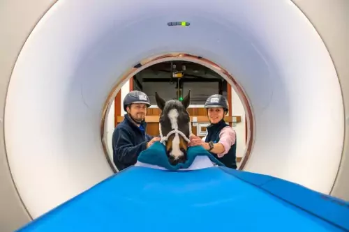
CT (Computed Tomography)
Computed tomography (CT) uses a rotating xray generator and detector plate to create cross sectional slices of the area being imaged. This removes the difficulties of superimposition of different structures that we have on xrays, giving clear images through the region. Assessment of soft tissues is also possible in addition to bone evaluation, and this can be improved with the help of contrast either into blood vessels, or directly into a joint or tendon sheath. CT with injection of contrast into a joint allows excellent assessment of the joint cartilage, and soft tissues within the joint.
At Bell Equine, we have two CT systems – the Qalibra fan-beam CT system and the Hallmarq Vision cone-beam CT.
Our state-of-of the art Qalibra CT system is one of only a few in the country that can image the lower limbs (up to and including the knee/hock) in addition to the head and upper neck in standing, sedated horses. The height of the scanner can be tailored to the size of the patient, allowing scanning of patients of all sizes, from Shetlands to Shires. Because of the large size of the bore (central hole) we are also able to image the whole spine, pelvis, thorax and upper limbs of adult horses (and foals/ponies!) under general anaesthesia.
The advantages of CT over x-rays for assessment of the horse’s head, especially sinus and dental disease are well known. However, with the ability to image more regions – lower limbs standing and upper limbs/neck under anaesthesia - CT has dramatically improved our ability to diagnose pathology of many regions, but particularly the stifle region, the neck, and the hock compared to traditional imaging (xrays and ultrasound).
With our Hallmarq Vision CT we can image the foot, pastern and fetlock in the standing, sedated horse. This is a cone-beam CT system which means the soft tissue assessment is much more limited than in the Qalibra system, but it can provide excellent bony detail. We run a Vision CT scan as part of the MRI package in horses undergoing MRI of the foot and fetlock, and it provides complementary information to the MRI scans.
Further Information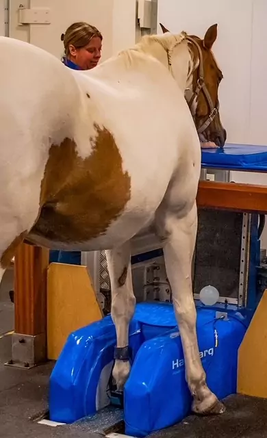
MRI
Bell Equine was the first equine clinic in the world to install an MRI (magnetic resonance imaging) scanner capable of imaging the limbs of a standing horse. The first scanner was installed here in 2002, and since then we have undertaken important evaluations of the system to continue to improve our image quality and scan higher up the limbs, and have collaborated with workers in a number of institutions around the world.
MRI allows the detailed examination of the different structures of the lower limb and is particularly useful in the evaluation of diseases of the foot. It yields in depth images of the anatomy including both the bone and the soft tissues. Images can be acquired in any plane to focus in on one area without superimposition of other structures. Our investigations have and continue to provide important and new information about many diseases of the foot, including the so-called "navicular disease". The procedure can also provide valuable information about more proximal areas of the limbs, such as the fetlock, knee and suspensory ligament.
After making sure there is no metal (or metal fragments from shoes or clenches) in the area to be imaged, the horse is taken to a specialised room for MRI. The room is shielded from all radio frequencies which would otherwise ruin the images. The area to be imaged is placed within the C-shaped magnet with the horse standing and a 'radiofrequency coil' which sends and receives signal, is placed around the limb. A variety of different sequences are obtained, each of which takes several minutes. Images are of the best quality if the horse stands still while each sequence is obtained, but the horse can move between sequences. Once all the images are obtained and the horse has recovered from the sedation, they are discharged from the clinic. The whole procedure normally takes approximately 4 hours but is variable depending on the area(s) imaged and how still we can keep the horse.
Further Information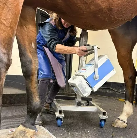
Radiography
The clinic currently has four X-ray generator units, including a powerful gantry-mounted unit. These allow us to obtain radiographs of high diagnostic quality of most parts of the horse’s body, including the upper limbs, neck and chest.
Our state-of-the-art digital radiography systems permit detailed image manipulation, thereby increasing the diagnostic value over conventional film-based radiography.
Ultrasonography
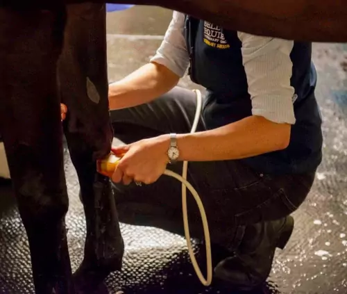
We currently have eight diagnostic ultrasound machines that have specialised probes and indications depending on which part of the horse we are imaging which allows a complete range of ultrasound examinations to be performed. These include probes and machines specifically designed for certain indications, ranging from small, high resolution imaging of tendons, ligaments, eyes and larynges, to deep penetrating imaging of hearts and intestines, as well as the ability to measure blood flow using doppler.
Ultrasound examinations are a major and valuable part of the investigation of horses affected by many orthopaedic diseases (e.g. tendon and ligament damage, joint evaluation, some fractures, back diseases, etc.), abdominal diseases (e.g. colics, abdominal tumours, weight loss cases, liver diseases, etc.), thoracic diseases (e.g. heart conditions, pleurisy, etc.), and gynaecological conditions (e.g. pregnancy diagnosis, pre-breeding checks and routine monitoring for A.I., ovarian abnormalities, etc.). Ultrasound guidance is commonly used for the precise and safe biopsy of the liver, kidney and other internal structures.
Nuclear Scintigraphy
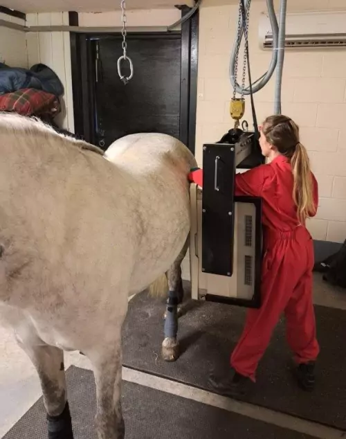
Nuclear scintigraphy, or bone scanning, is performed on over 100 horses per annum, usually as a part of a lameness evaluation, but also in some cases as a part of the evaluation of neurological or other conditions. This imaging modality gives an overview of bone turnover in the area imaged and is incredibly sensitive for detecting even subtle changes in bony turnover to help localise skeletal abnormalities.
We have a GE Medical Starport gamma camera mounted on an overhead gantry that permits imaging of the entire horse. The horse is injected with a special radioactive dye that binds to bone. After a few hours have passed allowing the dye to bind to the bone, the camera that detects the gamma radiation is used to scan the horse and generates images, thereby giving information about bone turnover. Horses undergoing this examination need to be hospitalised for at least 48 hours following an injection with the radioactive drug.